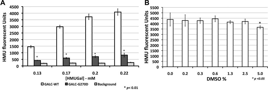
Figure 2. Development of GALC assay in multi-well plates.
Optimization of the assay was performed in multi-well plates. (A) Substrate concentration assays were performed using cultured SV40T skin fibroblasts in 384-well plates. The cells were cultured to full well-confluence and before starting the GALC assay. Substrate concentrations in the assay reaction over 0.2 mM of HMUGal generate elevated signal-to-background ratios (>5). GALC enzymatic activity from control cells with wild type (GALC-WT) produced higher HMU fluorescence signal (white bars). HMUGal substrate was able to detect a small residual activity of SV40T mutant GALC (GALC-G270D – dark gray columns) above the background, which represent fluorescence signals from wells with culture medium only (no cells). (B) The DMSO assay was performed in SV40T control cells (GALC-WT) demonstrated that GALC activity is reduced as DMSO concentration is increased in reaction solution. The DMSO concentrations of 2.5% or lower showed not to affect GALC enzymatic activity.

