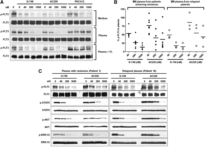
Figure 3.
Potent inhibition of G-749 in AML patient plasma milieu. (A) Molm-14 cells were treated with increasing concentrations of indicated inhibitors in normal culture medium (top), normal human plasma from healthy donor (middle), or normal plasma supplemented with 5 ng/mL of FL (bottom) for 2 hours. The autophosphorylation level of FLT3 was then evaluated in western blot. (B) The bone marrow (BM) plasmas were obtained from AML patients who achieved CR (n = 7) or those with relapse (n = 3) after the induction therapy of cytarabine. Molm-14 cells were treated with the indicated concentrations of G-749 or AC220 in relapsed or CR patient plasma milieu for 2 hours. The p-FLT3 was then analyzed by densitometry and plotted as the percentage over dimethylsulfoxide (DMSO) control to display distribution of data. ANOVA followed by Newman-Keuls multiple comparison test was performed to examine the inhibition level of p-FLT3. P < .05 was calculated between G-749 and AC220 in each concentration for pairwise comparison. Noticeably, even groups treated with 1000 nM AC220 showed greater deviation for inhibition levels of p-FLT3 from 10% to 100% residual p-FLT3 in both relapsed and CR plasma. G-749 showed less deviation for this than AC220 in all tested concentrations. (C) The phosphorylation levels of FLT3, STAT5, ERK1/2, and AKT were further evaluated in patient plasma milieu from the patient achieving remission (patient 1, left-side blot) and the relapsed patient (patient 10, right-side blot). Noticeably, G-749 potently inhibited FLT3 and downstream pathways in both remission and relapsed plasma, whereas AC220 significantly lost its potency against them in relapsed plasma.

