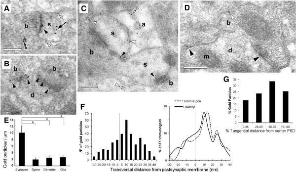
Figure 2.

Ultrastructural localization of ZnT1. Electron micrographs of Lowicryl sections showing ZnT1 particles (10 nm gold) at synapses made on dendritic spines (A, C) or shafts (B, D). Particles predominated over synaptic junctions (solid arrowheads). Extrasynaptic labeling was seen over the plasma membrane of spines and dendrites (white arrowheads). Occasional labeling on extrasynaptic membranes apposed to boutons (white arrow in D), inside astrocytes (double white arrowhead in C), inside spines (solid arrow in A), or over presynaptic vesicles (double solid arrowhead in A). b, bouton; s, spine; d, dendrite; a, astrocyte; m, mitochondrion. Scale bars, 0.2 μm. (E) Surface densities of ZnT1 (particles/μm) over different plasma membranes. (F) Left, Frequency distribution of gold particles across the synaptic junction (i.e. axodendritic). Negative and positive values denote locations pre- and post-synaptic to the outer leaflet of the postsynaptic membrane, respectively. Distances between the center of gold particle and membrane were grouped into 5 nm bins. Particles followed a normal distribution with a peak located at the +10 nm bin. Right, Relative distribution of ZnT1 across the synaptic junction analyzed in Lowicryl and Epon sections. (G) Relative distribution of ZnT1 along the PSD length. Particle location was computed as a fraction of the distance from the center (0%) to the edge (100%) of the PSD.
