Figure 3.
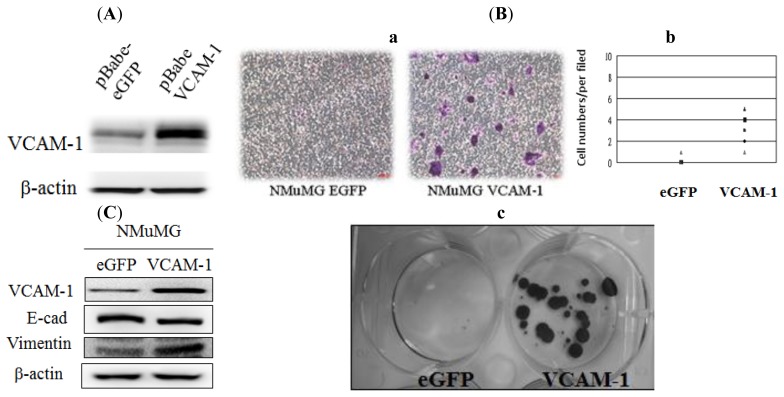
(A) Western blot VCAM-1 protein expression in NMuMG cells transfected with the pBabe-eGFP or the pBabe-VCAM-1 plasmid. β-actin results indicate similar sample loads; (B) a, Transwell migration analysis of NMuMG cells (control and VCAM-1 over-expression) towards 10% FBS + insulin. Photo images are representative fields of the migration of VCAM-1 over-expressing cells and the control group after crystal violet dye staining; b, The cell numbers of migrated NMuMG (eGFP control) and VCAM-1 NMuMG cells from the upper insert to the bottom surface of the lower well under a light micropore (100×) after overnight incubation; c, Migrated NMuMG VCAM-1 cells in the lower wells showed high clonogenic ability after 2 weeks of incubation; (C) EMT-related protein expression in eGFP control and VCAM-1 over-expressing NMuMG cells was determined by Western blot analysis.

