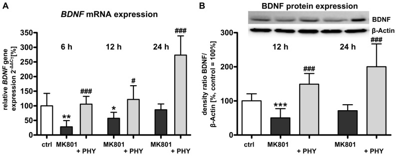
Figure 2.
Increased brain-derived neurotrophic factor (BDNF) expression after physostigmine co-treatment in MK801 treated rat pups. (A) Quantitative analysis of brain BDNF mRNA expression at 6, 12 and 24 h after MK801 treatment (dark grey bars) with or without systemic physostigmine co-application (light grey bars); and (B) Quantitative protein expression of BDNF at 12 and 24 h after MK801 treatment (dark grey bars) in combination with AChE inhibition (light grey bars). The densitometric data represent the ratio of the pixel intensities of BDNF signals to the corresponding β-actin signals. Data are normalized to levels of vehicle treated pups ((control; white bar, 100%); bars represent mean + SD, n = 6–7 per group, *** p < 0.001, ** p < 0.01, * p < 0.05 compared to vehicle treated pups, ### p < 0.001, # p < 0.05 compared to MK801 treated pups, respectively). ctrl: control; PHY: physostigmine.

