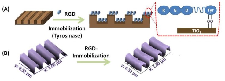
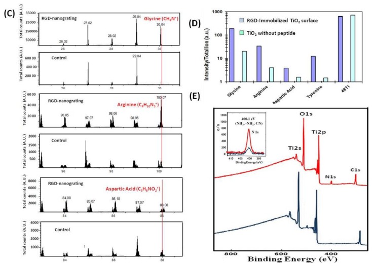
Figure 1.
(A) Schematic showing the attachment of RGD on TiO2 nanogratings; (B) 3-D AFM images showing retention of the TiO2 nanopatterns after RGD immobilization; (C) Time-of-Flight Secondary Ion Mass Spectrometry (ToF-SIMS) confirmed the attachment of RDG peptide on the TiO2 surface. The specific fragment pattern generated by removal of individual amino acids from peptide is shown in the lower panels. (Glycine m/z 30.04 u:CH4N+, Arginine m/z 100.07 u:C4H10N+, Aspartic acid m/z 88.08 u:C3H6NO2+); (D) Typical peak ratio of amino acids normalized by total ions; (E) XPS spectra; wide scan of N1s spectra of TiO2 surfaces before and after the immobilization of peptide. Inset: a high-resolution scan of N1s.


