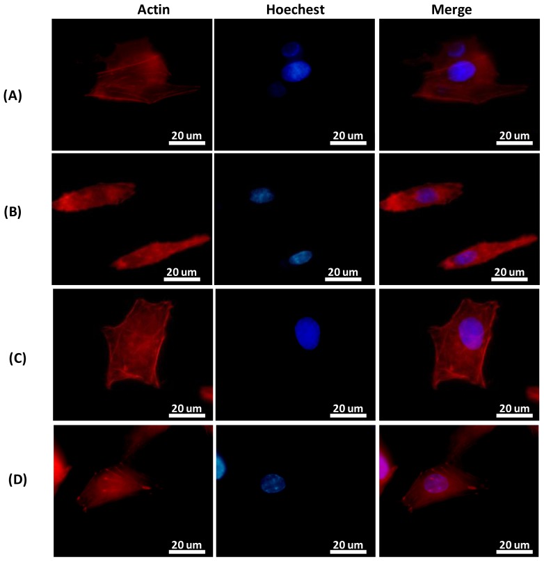
Figure 5.
Actin fluorescent staining to study the cell alignment and cytoskeletal rearrangement of HepG2 cells cultured for 12 h on (A) RGD-TiO2 nanogratings compared to (B) TiO2 nanopattern alone, on which the actin filaments are arranged parallel to the TiO2 nanofeatures (C) RGD-TiO2 flat surface and (D) Flat surface alone or control.

