Figure 7.
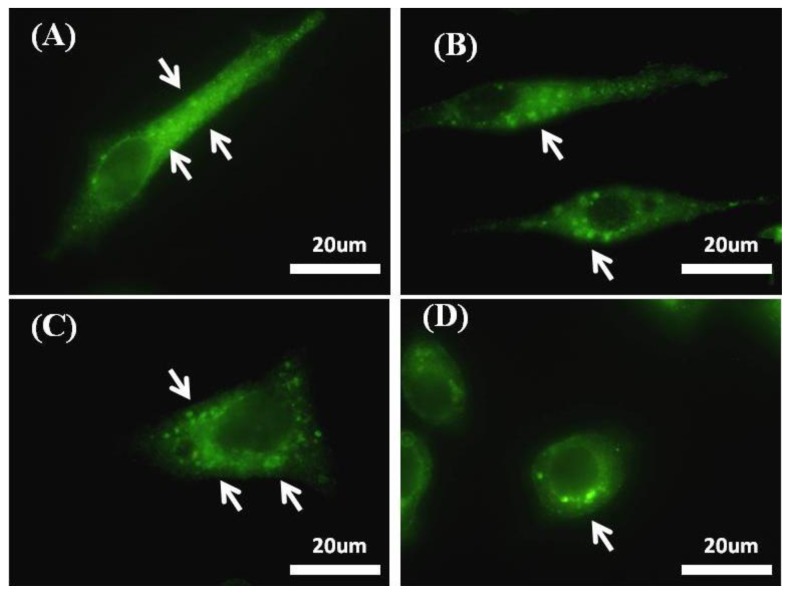
An increase in integrin β1 clusters that mediate focal adhesion formation using immunostaining of integrin B1 by Alexa-fluor 488 linked-AB (excitation λ = 488 nm) of HepG2 cells cultured on RGD-TiO2 substrates on which (A) RGD-nanogratings show high integrin β1 clusters while (B) Nanogratings alone show low integrin B1 clustering compared to it, on the other hand (C) RGD-flat surface shows high integrin β1 clusters, mean while (D) flat substrate alone (control) has the lowest integrin clustering; arrow heads show clusters of integrin B1 as green fluorescent dots.

