Figure 1.

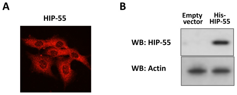
Expression and purification of HIP-55 protein. (A) Cells were fixed and labeled with antibodies to HIP-55. Subcellular location of HIP-55 was shown by laser scanning confocal microscopy; (B) After His-pull-down, the overexpression of HIP-55 in the HEK293 cell line was detected with Western blot assay by HIP-55 antibody. Actin was used as a loading control.
