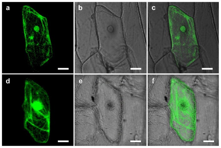
Figure 2.
Sub-cellular localization of CgHSP70 in transiently transformed onion epidermal cells. (a–c) Onion epidermal cells transiently expressing 35S::CgHSP70-GFP; (d–f) Onion epidermal cells transformed with a control construct (35S::GFP); (a,d) Dark field images to capture GFP fluorescence; (b,e) bright field images to capture cell features; (c,f) merged images. Scale bar: 25 μm.

