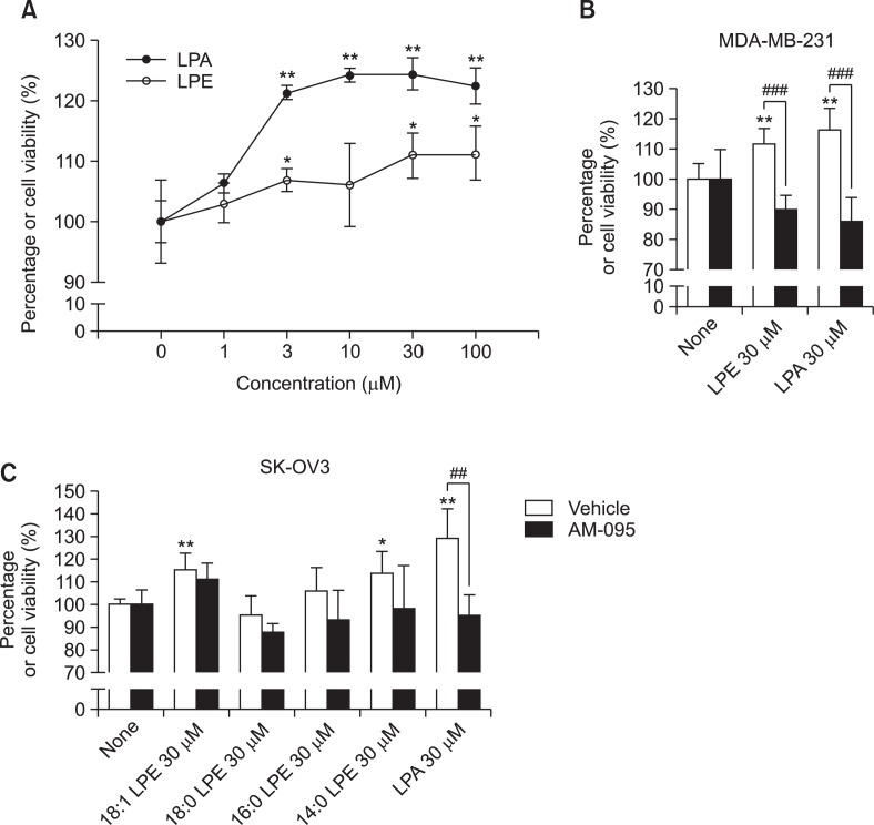
Fig. 3.
Effects of LPE and LPA on cell proliferation in MDA-MB-231 human breast cancer cells. MDA-MB-231 human breast cancer cells were treated with 0-100 μM 18:1 LPE or LPA for 24 h (A). Cell viabilities were measured using an MTT assay, as described in Materials and Methods. MDA-MB-231 human breast cancer cells were pretreated with 500 nM of AM-095 or vehicle for 5 min and treated with 30 μM of LPE or LPA for 24 h (B). SK-OV3 human ovarian cancer cells were pretreated with 500 nM of AM-095 or vehicle for 5 min and treated with 30 μM of LPE or LPA for 24 h (C). Results are the means ± SEs of three independent experiments. Statistical significance: *p<0.05, **p<0.01 vs. none-treated cells and ##p<0.01, ###p<0.001 vs. vehicle-treated cells.

