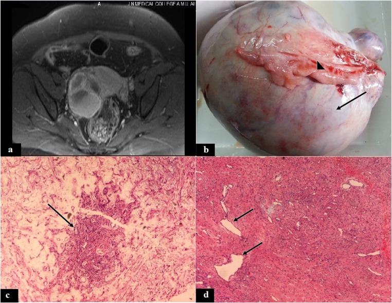
Figure 1.
(A) T2-weighted MRI showing a heterogeneous, solid-cystic tumour in the right adnexal region, (B) ovarian tumour with white surface (arrow) and attached fallopian tube (arrow head), (C) showing a tumour pseudolobule (arrow) with surrounding oedematous stroma (H&E, × 50), (D) pseudolobule with thin-walled hemangiopericytoma-like blood vessels (arrow) and fibrocollagenous stroma (H&E, ×50).

