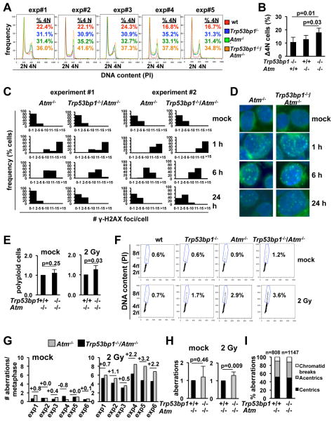
Fig. 4.
53BP1 deficiency increases the number of chromosomal translocations in irradiated Atm−/− primary cells. A, α-CD40/IL-4-activated B cells were irradiated, allowed to repair for 24 hours and stained with propidium iodide (PI) for analysis of cell cycle distribution. B, increase in the number of cells with 4N DNA content 24 hours after IR, relative to wt cells in the same experiment. Bars represent the average and standard deviation of the 5 experiments in A. C, distribution of the number of γ-H2AX foci per nucleus. N=100 cells per histogram. D, representative microphotographs of cells in C. E, frequency of cells with >4N DNA content 24 hr after IR. Data for the Trp53bp1−/−/Atm−/− culture was normalized to the Atm−/− control in the same experiment. Bars represent the average and standard deviation of 5 mice per genotype in 5 independent experiments. F, representative FACs plots of E. G, quantification of the number of chromosomal breaks 24 hours after IR using telomere FISH. H, the frequency of chromosomal breaks per metaphase in Trp53bp1−/−/Atm−/− cultures was normalized to Atm−/− cultures in the same experiment. Bars represent the average and standard deviation of 6 mice per genotype in 6 independent experiments. I, chromosomal breaks in G were classified as “chromosome-type” (centric and acentric chromosomes) or “chromatid-type”. The total number of aberrations analyzed is indicated.

