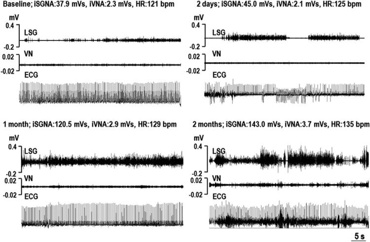
Figure 1. Examples of SGNA and VNA After MI.
Each panel shows the simultaneous recordings of the stellate ganglion nerve activity (SGNA), vagal nerve activity (VNA), and subcutaneous electrocardiogram (ECG) at around 11:00 am. The SGNA and VNA increased with increasing time from myocardial infarction (MI). bpm = beats per minute; HR = average heart rate; iSGNA = integrated stellate ganglion nerve activity over 1 min; iVNA = integrated vagal nerve activity over 1 min; LSG = left stellate ganglion; VN = vagal nerve.

