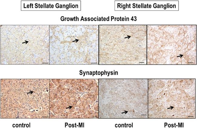
Figure 5. Immunohistochemical Staining of the Stellate Ganglia.
Immunohistochemical staining with growth-associated protein 43 (upper panel) and synaptophysin (lower panel) of left and right stellate ganglia in control dogs and in dogs with myocardial infarction (MI). Arrows point to positive stains (brown). The figures show that growth-associated protein 43–positive and synaptophysin-positive nerve structures are both more prominent in myocardial infarction dogs than in control dogs. The line segments are 50 μm long.

