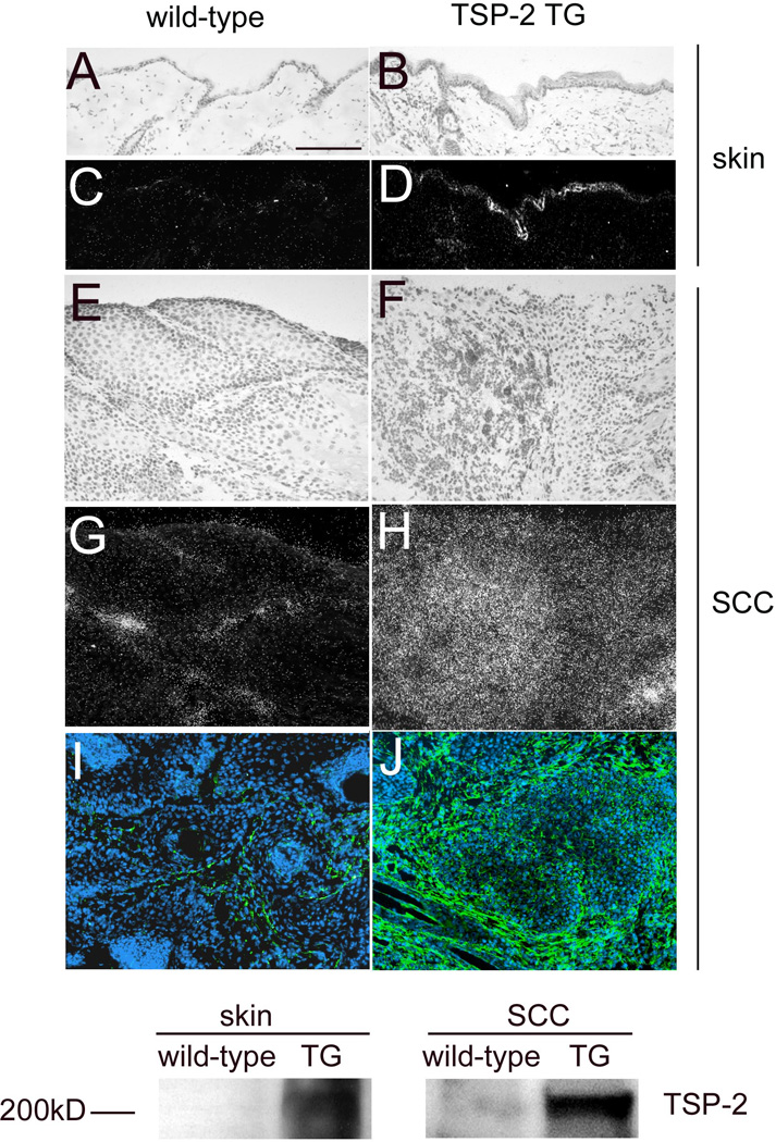
Figure 3. Differential TSP-2 expression in normal skin, SCC and tumor stroma in wild-type and transgenic mice.

In situ hybridization confirmed moderately enhanced TSP-2 mRNA expression in the normal skin (B,D) of TSP-2 transgenic mice and a strong TSP-2 expression in the SCC of TSP-2 transgenic mice (F, H), whereas little or no TSP-2 mRNA was detectable in the normal skin (A, C) or the SCC of wild-type mice (E, G). Immunofluorescence stains for TSP-2 (green) demonstrate pronounced deposition of TSP-2 protein predominantly in the tumor stroma in TSP-2 transgenic SCC (J), as compared to weak TSP-2 protein expression in wild-type SCC (I). Bar = 100 µm. Western blot demonstrates high levels of TSP-2 expression in the skin and in tumors of TSP-2 transgenic mice in contrast to low protein levels in wild-type mice (K).
