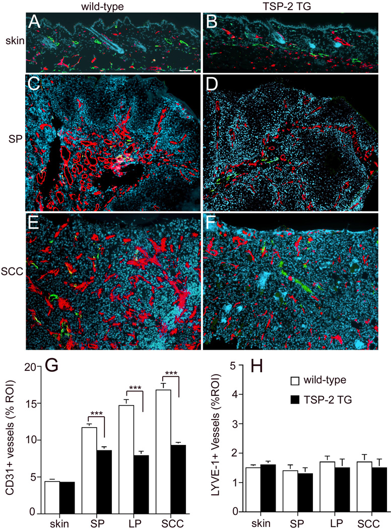
Figure 4. Diminished tumor angiogenesis in TSP-2 transgenic mice.
Differential immunofluorescence analyses of untreated skin for CD31 (red) and LYVE-1 (green) demonstrate comparable vascularization in wild-type (A) and TSP-2 transgenic skin (B). Increased density of CD31+ vessels in small papillomas (SP; C, D) and squamous cell carcinomas (SCC; E, F) in both genotypes is accompanied by insignificant alterations of LYVE-1+ vessels. Tumor angiogenesis was less pronounced in TSP-2 transgenic mice with a prominent reduction of enlarged angiogenic vessels. Bar = 100 µm. Quantitative image analysis of CD31-stained vessels revealed a significant decrease of the average vascular area in TSP-2 transgenic mice (filled bars) already at early stages of papilloma formation and throughout the progressive stages of skin carcinogenesis, as compared with wild-type mice (open bars, G). Quantitative image analysis of LYVE-1+ lymphatic vessels revealed a comparable average lymphatic vascular area in untreated skin and throughout the development of skin tumors in wild type (open bars) and TSP-2 transgenic mice (filled bars, H). skin; untreated skin (n=9), small papilloma (SP <3mm, n=11), large papilloma (LP >3mm, n=11), squamous cell carcinoma (SCC, n=11). Data are expressed as mean values+SEM., ***p<0.001.

