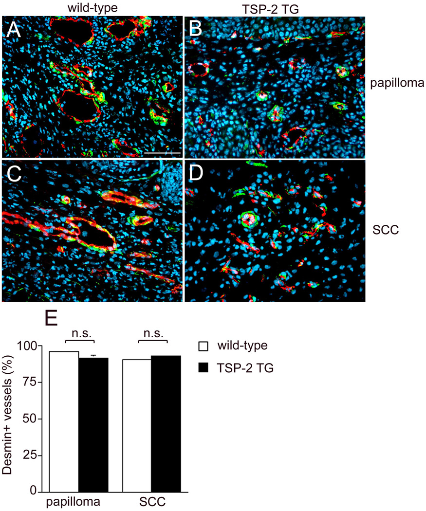
Figure 8. Comparable expression of desmin associated CD31-positive vessels both in wild-type and transgenic mice.
Differential immunofluorescence stains for CD31 and the pericyte marker desmin showed that the majority of vessels were surrounded by pericytes in papillomas (A, B) and SCC (C, D) of both genotypes. Bar = 50 µm. Quantification of desmin-associated CD31-positive vessels demonstrated a comparable percentage of desmin-positive vessels (E). Values expressed as mean+SEM; n.s.: not significant).

