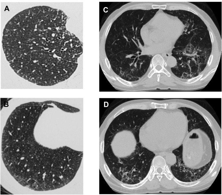
Figure 1.
High-resolution CT of the chest illustrating differences in the radiographic appearance of the lungs in the giant cell interstitial pneumonia (GIP) and the usual interstitial pneumonia (UIP) pattern. (A,B) In GIP of case 9, centrilobular micronodular opacities pathologically correspond to centrilobular fibrosis and giant cell accumulation within the alveolar space. (C,D) In the UIP pattern of case 10, reticular opacities and traction bronchiectasis are present with centrilobular micronodular opacities.

