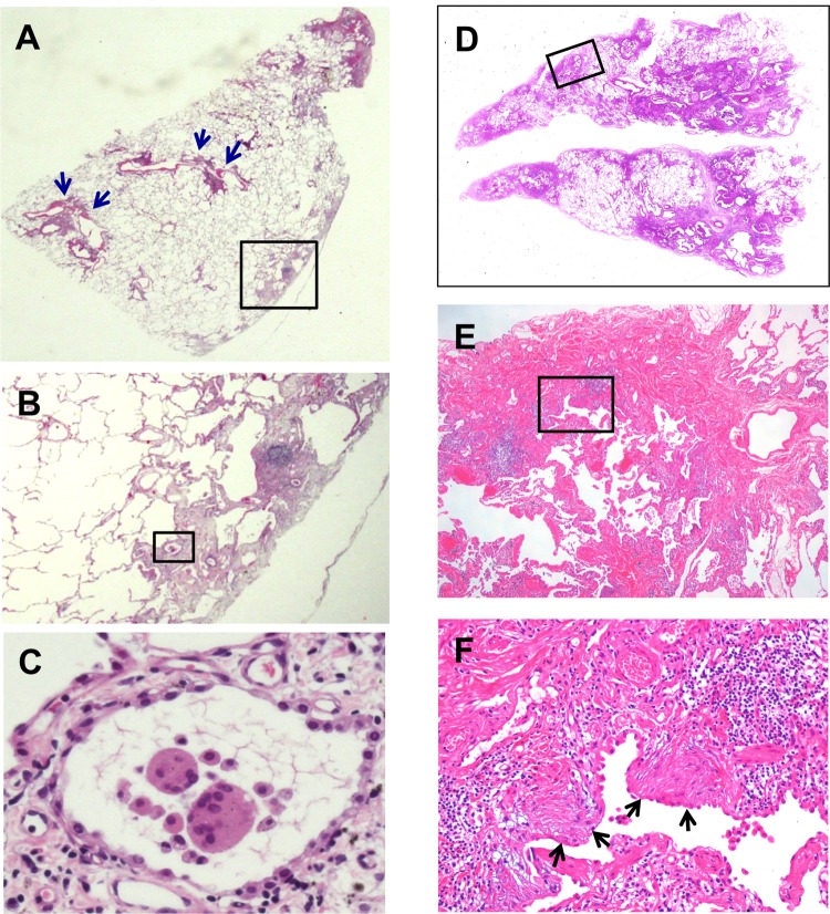
Figure 3.
Representative images of light microscopic findings of a lung specimen from case 10 with hard metal lung disease pathologically diagnosed as usual interstitial pneumonia pattern. (A,B) A low magnification view of the left S1+2 specimen demonstrates a combination of patchy interstitial fibrosis with alternating areas of normal lung and architectural alteration due to chronic scarring or honeycomb change. Note that there are several small bronchioles with mild centrilobular inflammation (blue arrows). (B,C) Multinucleated giant cells with cannibalism are also shown in a stepwise-magnified black square area located in subpleural fibrosis. (D–F) Left S10 specimen from the same patient also shows characteristic fibroblastic foci (black arrows) in the background of dense acellular collagen in a stepwise-magnified square area located in subpleural fibrosis. Original magnification, (A and D) panoramic view, (B) ×2, (C) ×40, (E) ×4, (F) ×20.

