Figure 2.
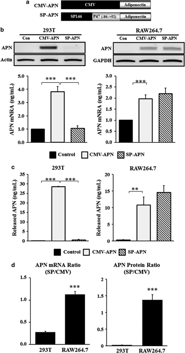
Constructs of plasmid vectors for APN expression. (a) SP146-C1 was selected to develop a therapeutic vector. The original luciferase gene in the pGL3-basic plasmid was replaced by the APN gene. (b) Cells were transfected and mRNA expression of APN with CMV-APN and SP146-C1-APN (SP-APN) was detected 24 h later by RT-PCR in 293 T and RAW264.7 cells. CMV-APN was used as a positive control. Densities of bands were measured using Alpha easy FC software and normalized to that obtained with actin or glyceraldehyde 3-phosphate dehydrogenase (GAPDH). (c) Concentrations of APN in the media were measured 48 h later using a human APN ELISA Kit in 293 T and RAW264.7 cells. Data shown are means±s.d.'s. (d) The ratio of expressions of SP-APN and CMV-APN was calculated and expressed in graphs. Both mRNA level of APN and released APN showed high ratio in RAW264.7 cells. **P<0.01 and ***P<0.001. APN, adiponectin; ELISA, enzyme-linked immunosorbent assay; GAPDH, glyceraldehyde 3-phosphate dehydrogenase.

