Figure 6.
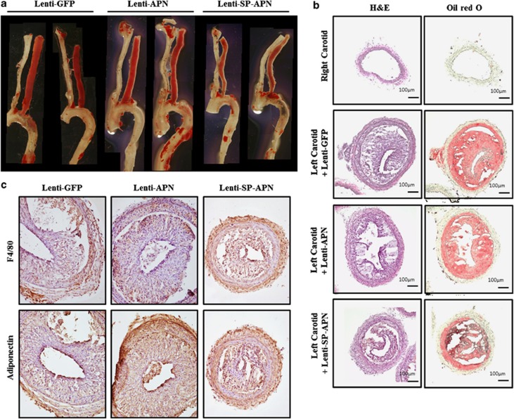
Macrophage-specific expression of APN reduced atherosclerotic lesions in ApoE knockout mice. (a) Representative images of Sudan-stained atherosclerotic lesions after application of lentiviral vectors for 2 weeks. Lenti-SP-APN-infected arteries showed a significant reduction of lipid deposit compared with that in Lenti-GFP- and Lenti-APN-infected arteries. (b) Hematoxylin and eosin (H&E) staining and oil red O staining were performed to characterize the lesions in lentiviral vector-infected lesions. (c) Macrophages were observed in all the lesions (F4/80). The expression of APN was detected in Lenti-APN- and Lenti-SP-APN-infected lesions. The size of the lesion was reduced by Lenti-SP-APN infection. APN, adiponectin.

