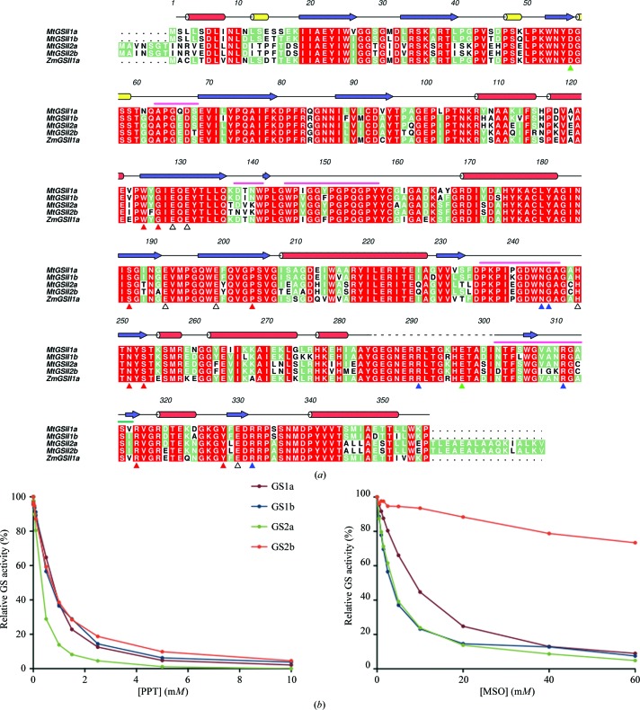
Figure 1.
(a) Amino-acid sequence alignment of plant glutamine synthetases, including M. truncatula GSII-1a (MtGSII1a; UniProt O04998), GSII-1b (MtGSII1b; UniProt O04999), GSII-2a (MtGSII2a; UniProt Q84UC1), GSII-2b (MtGSII2b; UniProt E1ANG4) and Z. mays GSII-1a (ZmGSII1a; PDB entry 2d3a; Unno et al., 2006 ▶; differing from UniProt B9TSW5 by a I353V replacement), performed with ClustalW (Larkin et al., 2007 ▶). The transit peptides of M. truncatula GSII-2a and GSII-2b were omitted. Red, green and white backgrounds are used for high, medium and low conservation, respectively. Numbers above the alignment and secondary-structure elements (α-helices in red, 310-helices in yellow and β-strands in blue) correspond to the crystal structure of MtGSII-1a described here. A dashed line denotes disordered residues in the crystallographic model. The ligand-contacting residues in Z. mays GSII-1a are indicated below the alignment with red (ATP), blue (glutamate), green (ammonia) or open (metal coordination) triangles. A pink line above the alignment indicates the structurally distinct regions between MtGSII-1a and ZmGSII-1a. Prepared with Aline (Bond & Schüttelkopf, 2009 ▶). (b) Inhibition of M. truncatula GSII by increasing concentrations of PPT (left) and MSO (right). The GS activity was normalized to that found in the absence of inhibitor. PPT and MSO were titrated using standard synthetase reactions with 100 or 20 mM glutamate, respectively.

