Figure 5.
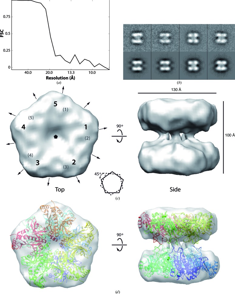
Electron cryomicroscopy reconstruction of M. truncatula GSII-2a. (a) FSC showing a resolution of 20 Å. (b) Pairs of class averages (upper row) and projections of the final three-dimensional reconstruction (bottom row). (c) Structure of M. truncatula GSII-2a, with approximate dimensions and positions of the symmetry axes (arrowed lines and pentagon for twofold and fivefold axes, respectively) defining the D5 symmetry. The numbers 1–5 and the numbers in parentheses indicate the positions of the five high-density blobs in the top and bottom rings, respectively. A scheme shows the relative rotation of the pentagons defined by the rings. (d) Fitting of the crystal structure of M. truncatula GSII-1a reported here, in which each monomer is coloured differently.

