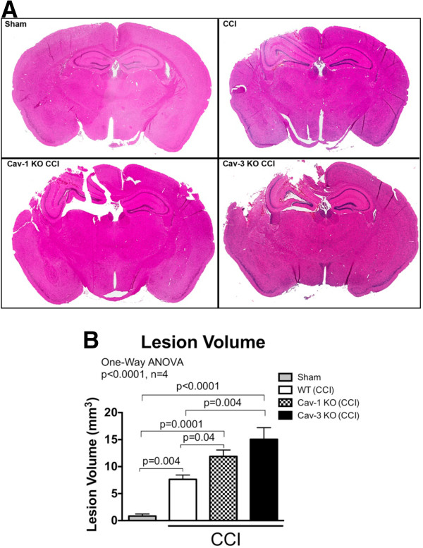
Figure 5.
Controlled cortical impact (CCI) causes a significant larger brain lesion volume in caveolin (Cav)-1 and Cav-3 knock-out (KO) mice compared wild-type (WT). WT, Cav-1 and Cav-3 KO mice were subjected to CCI and lesion volume was quantitated on Masson’s trichrome stained histological sections 24 hours following impact as previously described [22]. Cav-1 (11.9 ± 1.2 mm3) and Cav-3 KO (15.1 ± 2.2 mm3) mice had a significant larger brain lesion volume compared to WT (7.5 ± 0.8 mm3) and sham (0.8 ± 0.4 mm3) (P < 0.0001, n = 4). Data displayed as mean ± SEM. (A) Representative Masson’s trichrome stained coronal brain sections. (B) Quantitation of lesion volume shown in (A).

