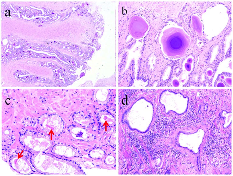
Figure 3.
Histopathology of prostatic lesions. (a) H&E staining (magnification, ×12.5) shows multiple corpora amylacea at the prostatic urethra. (b) H&E staining (magnification, ×100) shows corpora amylacea of the layer structure. They are frequently observed in benign glands but are rarely noted in carcinomas. (c) H&E staining (magnification, ×100) shows prostatic crystalloids (arrows). They are often observed in malignant or atypical glands. (d) H&E staining (magnification, ×40) shows prostatitis with moderate lymphocyte infiltration.

