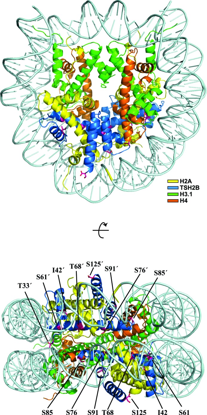
Figure 2.
Crystal structure of the TSH2B nucleosome. Two views are presented, in which the TSH2B molecules are coloured blue and the H2A, H3.1 and H4 molecules are coloured yellow, green and orange, respectively. The TSH2B-specific residues are indicated. Dyad symmetry-related residues are denoted by a prime (′).

