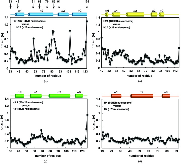
Figure 3.
Structural differences between the histones in the TSH2B and canonical H2B nucleosomes. (a) The TSH2B and H2B structures were superimposed and the r.m.s.d. value for each residue pair was plotted. The secondary structure of TSH2B in the nucleosome is shown at the top of the panel. Arrows indicate the locations of the TSH2B-specific amino-acid residues. (b) The H2A structures of the TSH2B and H2B nucleosomes were superimposed and the r.m.s.d. value for each residue pair was plotted. The secondary structure of H2A in the nucleosome is shown at the top of the panel. (c) The H3.1 structures of the TSH2B and H2B nucleosomes were superimposed, and the r.m.s.d. value for each residue pair was plotted. The secondary structure of H3.1 in the nucleosome is shown at the top of the panel. (d) The H4 structures of the TSH2B and H2B nucleosomes were superimposed, and the r.m.s.d. value for each residue pair was plotted. The secondary structure of H4 in the nucleosome is shown at the top of the panel.

