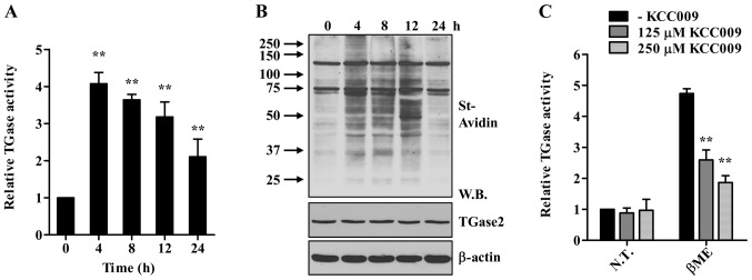
Figure 1.

Treatment with β-ME activates TGase2 in HLE-B3 cells. (A and B) HLE-B3 cells were exposed to β-ME (7.5 mM) for the indicated times. Cells were incubated for 1 h with BP (1 mM), and intracellular TGase2 activity was determined by microtiter plate assay (A) and western blot analysis (B). Streptavidin-HRP (St-Avidin) was used to detect the BP incorporated into the proteins. **p<0.01 compared to 0 h. (C) In situ TGase2 activity in HLE-B3 cells exposed to β-ME for 4 h in the absence or presence of the indicated concentrations of KCC009. Relative TGase2 activity is expressed as the fold-change compared with the values for non-treated cells. Results are presented as means ± SD (n=3). **p<0.01 compared to cells in the absence of KCC009 (two-way ANOVA with Bonferroni post-test). β-ME, β-mercaptoethanol; BP, biotinylated pentylamine; W.B., western blot; N.T., not treated.
