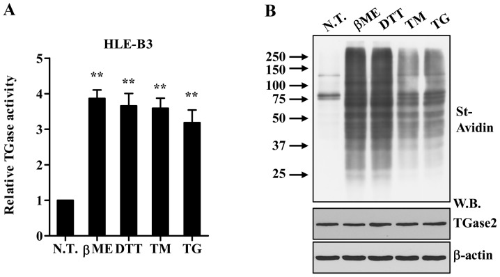
Figure 2.
Various ER stress-causing agents activate TGase2 in HLE-B3 cells. (A and B) In situ TGase2 activity in HLE-B3 cells exposed to β-ME (7.5 mM) or DTT (3 mM) for 4 h and TG (1 mM) or TM (5 μg/ml) for 24 h. Enzymatic activity was determined by microtiter plate assay (A) and western blot analysis (B). **p<0.01 compared to N.T. N.T., not treated; β-ME, β-mercaptoethanol; DTT, dithiothreitol; TM, tunicamycin; TG, thapsigargin; W.B. western blot.

