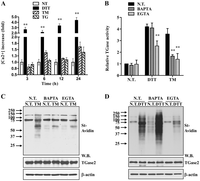
Figure 3.
ER stress activates TGase2 through an increase in intracellular calcium. (A) Increase in [Ca2+]i in response to ER stress. Data are expressed as the means ± SD (n=6). **p<0.01 compared to N.T. (two-way ANOVA with Bonferroni post-test). (B) Intracellular TGase2 activity was measured in HLE-B3 cells following exposure to 5 μg/ml TM or DTT for 24 h in the presence or absence of either EGTA (1.5 mM) or BAPTA-AM (20 μM). (C and D) Equal amounts of whole cell extracts were immunoblotted with HRP-conjugated streptavidin (St-Avidin) (upper panel) and antibodies specific for TGase2 and β-actin, respectively (lower panel). N.T., not treated; DTT, dithiothreitol; TM, tunicamycin; TG, thapsigargin; W.B., western blot.

