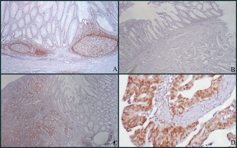
Figure 1.
(A) There is no staining in non-neoplastic surface epithelium, whereas strong nuclear staining is observed in lymphoid follicles (HMBG1 ×100). (B) Staining score 0 (negative): There is no staining in non-neoplastic surface epithelium and tumor cells (HMBG1 ×100). (C) Staining score 5 (positive): No staining is observed in non-neoplastic surface epithelium, but adenocarcinoma is stained diffusely and is moderately positive (HMBG1 ×100). (D) Staining score 6 (positive): There is diffuse and strong nuclear staining in adenocarcinoma and accompanying weakly positive cytoplasmic staining is observed (HMBG1 ×200).

