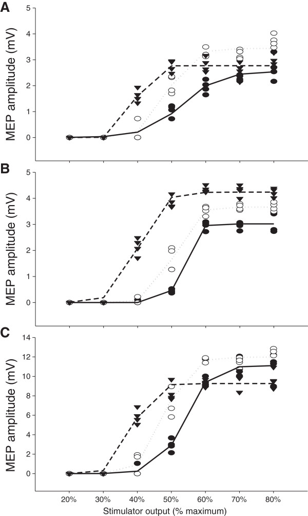
Figure 4.
Sample Boltzmann curves. Boltzmann sigmoidal function plotted versus stimulator intensity for one subject for vastus lateralis in Panel A, rectus femoris in Panel B and vastus medialis in Panel C. All motor-evoked potentials used in the modeling and the Boltzmann curves are presented for stimulus–response curves at 10 (●, ), 20 (○,
  ) and 50% (▼,
  ) of maximal voluntary force.

