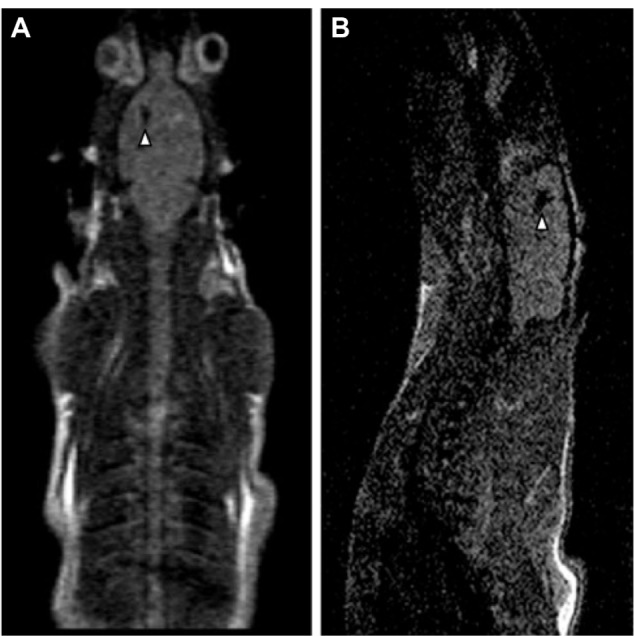
Figure 3.

Magnetic resonance imaging (MRI) of rats with neurodegenerative disease injected by mesenchymal stem cells labeled with magnetic nanoparticles.
Notes: In both MRI images (sagittal A and horizontal B) a dark area, identified by a white arrow, is present. This indicates a large concentration of SPION-labeled mesenchymal stem cells. Reprinted from Stem Cell Res, 9(2), Moraes L, Vasconcelos-dos-Santos A, Santana FC, et al, Neuroprotective effects and magnetic resonance imaging of mesenchymal stem cells labeled with SPION in a rat model of Huntington’s disease, 143–155, Copyright (2012), with permission from Elsevier.21
