Figure 3.
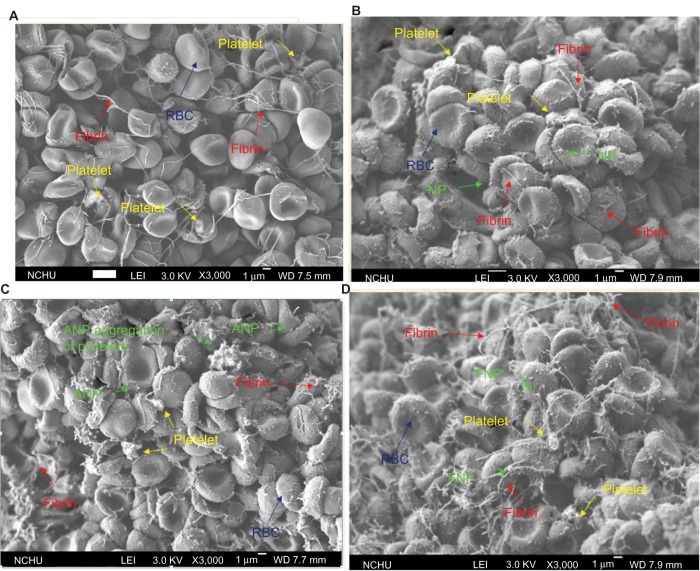
SEM micrographs of (A) clot/PBS showing the RBC aggregates, platelets, and fibrin network; (B) clot/NP showing many CS NPs adhering to the surfaces of RBCs or RBC aggregates; (C) clot/ANP showing many platelet aggregates in the clot; and (D) clot/FNP showing many FNPs adhering to the RBCs or RBC aggregates.
Note: In (D), the fibrin network is also much denser than that in other clots (B and C).
Abbreviations: ANP, adenosine diphosphate–decorated chitosan nanoparticle; Clot/ANP, clot induced in ANP solution; Clot/FNP, clot induced in FNP solution; Clot/NP, clot induced in CS NP solution; Clot/PBS, clot induced in phosphate-buffered solution; CS NP, chitosan nanoparticle; FNP, fibrinogen-decorated chitosan nanoparticle; RBC, red blood cell; SEM, scanning electron microscopy.

