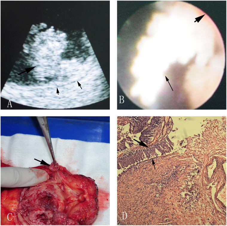
Figure 3. A nonmuscle-invasive bladder tumor near the bladder neck in ultrasonic image (A), direct version (B), radical cystectomy specimen (C) and pathological image (D).
A: The big black arrow indicates bladder tumor, and the two small arrows indicate continuous muscle layer; B: The small black arrow points at the bladder tumor, and the big one points at the bladder wall; C: The arrow shows us a bladder tumor near the bladder neck; D: The big black arrow indicates bladder tumor, and the small arrow indicates normal bladder epithelium.

