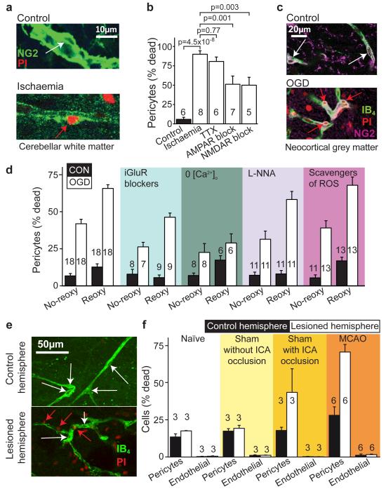
Figure 5. Pericyte death in ischaemia.
a Rat cerebellar slice white matter capillaries labelled for NG2 and propidium iodide (PI) after 1 hour of control (white arrow: living pericyte) or ischaemia solution containing antimycin and iodoacetate (red arrow: dead pericyte). b Percentage of pericytes dead in control or after 1 hour’s ischaemia (as in a) alone or with block of action potentials (TTX, 1μM), AMPA/kainate receptors (25μM NBQX), or NMDA receptors (50μM D-AP5, 50μM MK-801, 100μM 7-chlorokynurenate); p values from one-way ANOVA with Dunnett’s post-hoc tests. c Rat neocortical slice grey matter capillaries labelled for IB4, NG2 and PI after 1 hour’s control solution or oxygen+glucose deprivation (OGD). d Percentage of pericytes (as in c) dead after one hour’s OGD (No-reoxy) or OGD followed by 1 hour of control solution (Reoxy) with no drugs, or with iGluR block (NBQX (25μM), AP5 (50μM) and 7CK (100μM)), zero [Ca2+]o, NOS block (100μM L-NNA), or free radical scavenging (150μM MnTBAP or 100μM PBN, pooled data from Ext. Data Fig. 5a) throughout. OGD killed pericytes (ANOVA, p=10−13) and death increased during reperfusion (p=3.3×10⁻¹³). iGluR block or zero [Ca2+]o reduced death (ANOVA with Dunnett’s post-hoc test, p=2.7×10⁻⁴ and 6.0×10⁻⁷). Blocking NOS had a small protective effect (p=0.026); ROS scavenging did not (p=0.99). e-f Confocal images of striatal capillaries labelled with IB4 and PI (e) and percentage of striatal pericytes and endothelial cells that are dead (f) from the control and lesioned hemisphere of in vivo MCAO-treated rats (90 mins, assessed 22.5 hours later), sham-operated rats (with or without filament being inserted into the internal carotid artery (ICA)), and naïve control animals. More pericytes die than endothelial cells (repeated measures ANOVA, p=10−6). For pericytes, but not endothelial cells, cell death is greater in lesioned hemisphere (main effect of hemisphere, p=0.004; hemisphere-cell type interaction p=0.003) and cell death is greater in MCAO-lesioned animals than in naïve or sham animals without ICA occlusion (Tukey post-hoc tests, p=0.005 and 0.01). See Ext. Data Fig. 5 for data from cortex.

