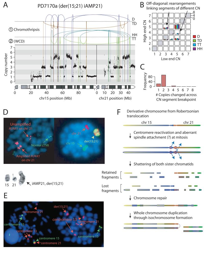
Figure 2. Rearrangements of der(15;21) in patient PD7170a.
A: Rearrangement and copy number pattern. The temporal order of the two major rearrangement events are marked ① and ②. Rearrangements are separated based on their orientation: D, deletion-type; TD, tandem duplication-type; HH, head-to-head inverted; TT, tail-to-tail inverted. B: Copy number jump distribution, showing the copy number at each end of each rearrangement. C: copy number step distribution, showing the distribution in magnitude of copy number change at copy number segmentation breakpoints. D: Representative metaphases: Left-hand cell shows multiple signals for RUNX1 (large red signals) clustered on two regions on the abnormal chromosome. And normal copies of ETV6 on chromosome 12 (green). Right-hand cell has been painted for chromosomes 15 (green) and 21 (red). Inset shows partial G-banded karyotype: normal chromosome 15, normal chromosome 21 and isochromosome der(15;21). E: Left-hand cell shows representative metaphase from a non-leukaemic cell in patient PD10009a with rob(15;21)c, hybridized with centromere-specific probes for chromosomes 15 (green) and 13 and 21 (red), confirming that the Robertsonian chromosome is dicentric. Right-hand cell shows a leukaemia metaphase in which der(15;21) iAMP21 chromosome retains the chromosome 21 centromere (red), but not the chromosome 15 centromere (green). F: Model for evolution of iAMP21 in rob(15;21)c. Newly synthesized sister chromatids are indicated by a blue outline.

