
Figure 2. Comparison of phenotype between CIK and DC-CIK cells on day28.
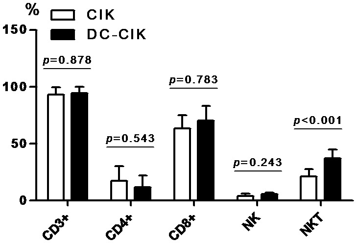
Compared with CIK cells, NKT cells were significantly increased in DC-CIK cells. However, there was no significant difference in the expression of CD3+, CD4+, CD8+ T cells and NK cells between CIK cells and DC-CIK cells.