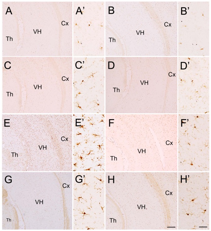
Figure 6. Iba1-immunopositive cells in VH, Cx and Th of saline- and LPS-injected rats.
A small number of Iba1-immunopositive cells are present in VH, Cx and Th of rats received neonatal intrahippocampal injection of saline (A) and the saline-injected rats treated with minocycline (B), risperidone (C) or both of them (D). On the other hand, a large of number of Iba1-immunopositive cells are observed in VH, Cx and Th of the rats received neonatal intrahippocampal injection of LPS (E), but it is dramatically reduced after intragastric administration of minocycline (F), risperidone (G) or both of them (H). A'–H' are high magnification of the ventral hippocampus in A–H, respectively. Cx, cerebral cortex; Th, thalamus; VH, ventral hippocampus. Scale bars = 200 μm in H (applied from A–G) and 20 μm in H' (applied for A'–G').

