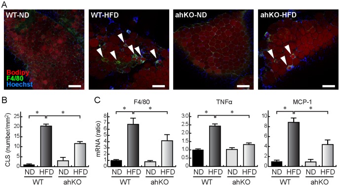
Figure 2. Inflammation of epididymal adipose tissues.
(A) Immunostaining for F4/80. Isolated epididymal fat pads were immunostained with anti-F4/80 antibody (green). Adipocytes were counterstained with BODIPY (red) and nuclei, with Hoechst (blue). Bars indicate 200 μm. Arrowheads indicate CLS. (B) The number of CLS in a 1 mm2 visual field was counted. Values are means ±S.E.M. (n = 6). *P<0.01. (C) Real-time PCR analyses of inflammation-related genes in epididymal adipose tissues. Values are means ±S.E.M. (n = 6). *P<0.05.

