Figure 2.
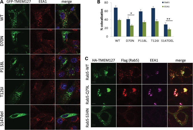
TMEM127 mutants have reduced association with the early endosome. (A) Confocal microscopy of GFP-fusion constructs encoding each of the four mutants or WT TMEM127 (green) co-stained with the early endosomal marker EEA1 (red). Merged images are shown in yellow and nuclei are DAPI stained (blue). Scale bars are 10 µm. (B) Degree of colocalization of each construct shown in (a) with EEA1 or with Rab5 (images shown in Supplementary Material, Fig. S1). Data were quantified from at least three independent experiments with at least 14 cells analyzed for each construct (*P < 0.05; **P < 0.01). (C) HA-TMEM127 (green) co-transfected with Flag-tagged Rab5 constructs WT, Q79L or S34N (red) and stained with endogenous EEA1 (magenta). Merged signals are shown in white. TMEM127 colocalization with the early endosomal marker EEA1 is increased by Rab5-Q79L expression and decreased by Rab5S34N. Note the lack of EEA1 staining in Rab5-positive structures on the bottom panel, indicating dysfunctional early endosomal assembly in Rab5S34N-transfected cells. Scale bars are 5 µm.

