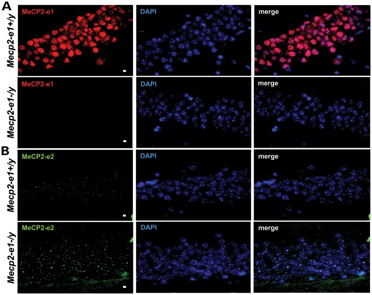
Figure 4.
MeCP2-e1 is ablated in Mecp2-e1−/y mouse brain, while MeCP2-e2 levels are elevated as shown by IF. (A). IF detection of MeCP2-e1 in mouse hippocampus using a MeCP2-e1 JL specific antibody in adult Mecp2-e1−/y mutant and Mecp2-e1+/y control brain sections. Alexa 594-tagged (red) secondary antibodies were used to visualize the distribution of MeCP2-e1 in the nucleus. Higher magnification images are shown in Supplementary Material, Figure S1. (B). IF detection of MeCP2-e2 in mouse hippocampus using an MeCP2-e2 antibody with Alexa 488 (green) conjugated secondary in Mecp2-e1−/y and WT Mecp2-e1+/y control hippocampal sections. DAPI staining as shown in blue reveals the location of the nucleus in all panels. The scale bar represents 5 microns.

