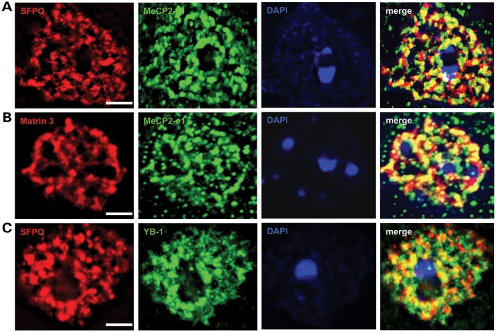
Figure 7.
MeCP2-e1 co-localizes with multiple nuclear matrix factors. (A) IF staining in cortical neurons for Matrin 3 (red) shows co-localization with MeCP2-e1 (green) distribution as shown by overlapping signal (yellow) in the merge panel. (B) Co-staining for SFPQ (red) and MeCP2-e1 (green) similarly shows significant signal overlap (yellow) indicative of co-localization in neuronal nuclei. (C). As a positive control, a previously identified MeCP2-associated factor YB-1 (red) also associates with the nuclear matrix as represented by SFPQ signal (green). Overlap of signal distribution is shown in the merged image. In all panels, the location of the nucleus is shown by DAPI counter staining (blue). Scale bars correspond to 5 μm.

