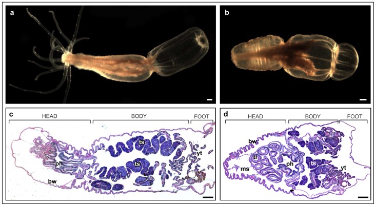
Figure 1. Nematostella vectensis morphology.
(a) five months old Nemtostella polyp in open position with extended tentacles. (b) five months old Nemtostella polyp in closed position with folded tentacles inside the pharynx. (c) H&E staining of a longitudal section of Nemtostella polyp in open position. No tentacle tissue present. (d) H&E staining of a longitudal section of Nemtostella polyp in closed position. Tentacle cross-sections appear inside the pharynx cavity. Scale bars: 200 μm.

