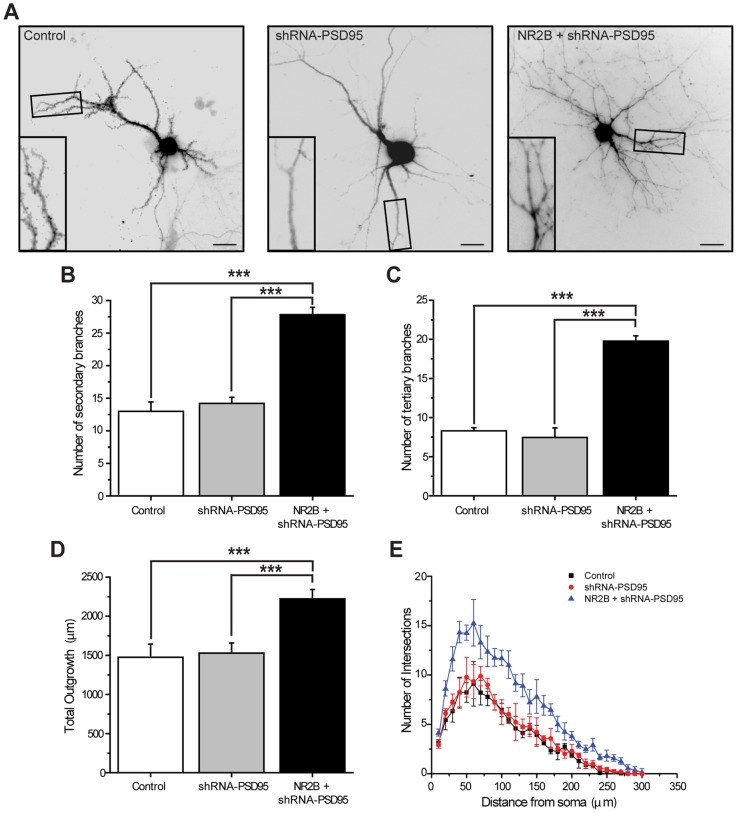
Figure 5. Simultaneous over-expression of NR2B and knockdown of PSD95 induce dendritic branching in mature hippocampal neurons.
A, Cultured hippocampal neurons were transfected with a magnetofection-based method at 15 DIV with GFP alone (control; left image), GFP plus shRNA-PSD95 (shRNA-PSD95; middle image), or GFP plus NR2B and shRNA-PSD95 (NR2B+shRNA-PSD95; right image). At 20 DIV, cultures were fixed and images taken. Scale bar is 25 μm. Inset: Magnified views of boxed areas showing examples of dendritic branches. Note that neurons that express NR2B+shRNA-PSD95 display a more complex dendritic architecture compared to control or shRNA-PSD95-expressing cells. B–C, Quantification of the average number of secondary (B) and tertiary (C ) processes: neurons expressing NR2B+shRNA-PSD95 have more branches relative to control neurons and to cells transfected with shRNA-PSD95. D–E, Total outgrowth (D) and Sholl analysis (E) of hippocampal neurons expressing the different constructs as indicated. For each condition, at least 20 neurons, obtained from 3 independent experiments, were analyzed. Figures show Mean ± SEM. *** p<0.001 (ANOVA).

