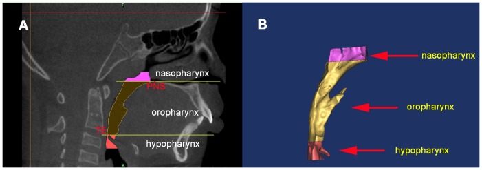
Figure 2. 3D model of the upper airway was reconstructed.

A) The upper airway was subdivided into three parts by two planes perpendicular to the sagittal plane and each region was highlighted in different colors. The landmarks used for defining the planes were: PNS, posterior nasal spine; SE, the superior border of the epiglottis. B) Each region of the upper airway was reconstructed respectively. The nasopharynx is the region from the top of the upper airway to PNS. Oropharynx region is between PNS and SE, and the hypopharynx is from SE to the level of the third cervical vertebra (C3).
