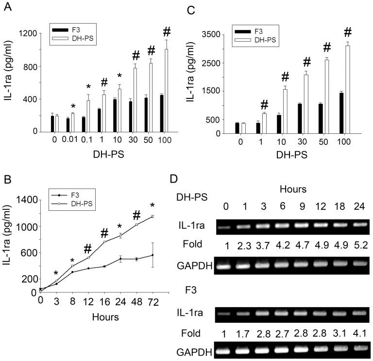
Figure 9. DH-PS induced more IL-1ra production than F3 in human CD14+ cells and THP-1 cells.
(A) Human CD14+ cells isolated from one healthy donor were cultured (2×10⁶ cells/ml) with increasing concentrations of DH-PS or F3 for 18 hours and supernatants were collected for the measurements of IL-1ra. (B) Human CD14+ cells were cultured with DH-PS (100 μg/ml) or F3 (100 μg/ml) and supernatants were collected at the indicated time points for IL-1ra measurements. (C) THP-1 cells were cultured (2×10⁶ cells/ml) with increasing concentrations of DH-PS or F3 for 18 hours and supernatants were collected for the measurements of IL-1ra. In A and C, X-axis represented the concentration of DH-PS (μg/ml). Concentration 0 represented the use of PBS only as vehicle control. Results were presented as mean concentrations of IL-1ra with error bars showing the standard deviation of triplicate. Statistically significant difference: * compared with F3-treated group, p<0.05. # compared with F3-treated group, p<0.005. (D) THP-1 cells were cultured with DH-PS (100 μg/ml) or F3 (100 μg/ml) and cells were collected at the indicated time points for the assessment of mRNA expression of IL-1ra.

