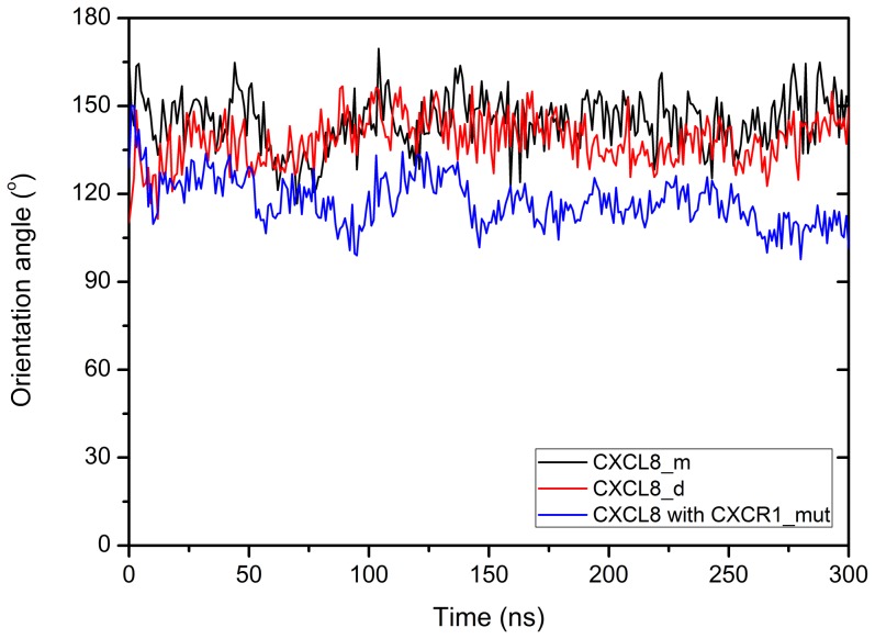
Figure 4. The orientation angle distribution of ligands binding with the receptor throughout the MD simulations.
The orientation angle of the bound ligand is defined as the angle between the unit vector normal to the membrane and the unit vector along the dipole of ligand. The averaged curves for three replicates of each system are shown for monomeric CXCL-8 in black, dimeric CXCL-8 in red, and CXCR1_mut in blue, respectively. Error bars of the curves are omitted for figure clarity.

