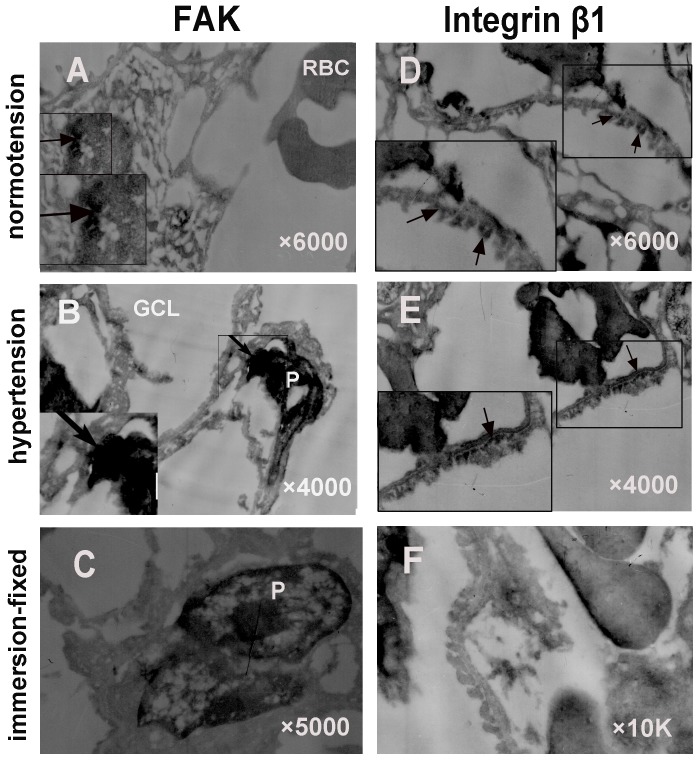
Figure 4. Immune electron micrographs of integrin β1 and FAK in glomeruli under normotensive and acute hypertensive conditions.
Under normotensive condition, the foot processes of podocytes were shown to be shrunken with the conventional fixation method (F). In contrast, using IVCT, the foot processes tightly approached each other and became flatter, integrin β1 was distributed on the basolateral membrane of podocytes (D), while FAK was located in the cytoplasm and nuclei of the podocytes (A). In contrast, under hypertensive condition, integrin β1 attached along the basal membrane only (E), while FAK was strikingly gathered in the nuclei (B). Left bottom pictures are the manified micrographs. They were manified 10 K. RBC: red blood cell; GCL; glomerular capillary loop; P: podocyte. (Magnification, ×6000 for A and D; ×4000 for B and E; ×5000 for C; ×10 K for F).

